Welcome to the latest edition of the BVNS Neurotransmitter.
Ziva’s Story
Martin Young, DVM, DACVIM (Neurology)
Ziva’s History
Ziva was adopted by her family at 7-years-old. Her background medical history includes coxofemoral osteoarthritis and previous treatment for heartworms. She presented to Bush Veterinary Neurology Service in Richmond, VA due to an acute onset of seizure activity. Her guardians noted that Ziva had her first seizure in mid November, 2021. She had facial twitching and went into a tonic clonic seizure that lasted 2-3 minutes. A couple of days later, she presented to her primary care veterinarian for bloodwork which came back within normal limits. Almost a month later, Ziva had her second seizure.
MRI
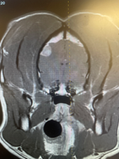
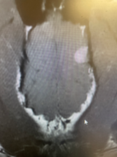
Dr. Martin Young at Bush Veterinary Neurology Service recommended and performed diagnostics including MRI of the brain. T1 post contrast sequences show an increased signal mass lesion within the left parietal lobe. The mass measured 0.8cm X 1cm.
Summary: these findings are consistent with a small left forebrain meningioma.
 Coronal T1 post contrast Coronal T1 post contrast |
 Axial T1 post contrast Axial T1 post contrast |
Prognosis & Treatment
Based on the diagnostics, the prognosis for Ziva is fair to good depending on the treatment course. With medical management of steroids and an anticonvulsant medication, there would be an anticipated survival up to 6 months, with the addition of chemotherapy, about 8-10 months, and with radiation therapy, there would be an expected survival of 14-18 months. If decompressive surgery is considered, survival rate of over 18 months is expected. A combination of therapy would anticipate a longer survival which has been reported out to 3 years. After the MRI diagnosis, Ziva started a steroid medication to reduce edema and inflammation and an anti-convulsant medication to manage seizures prior to decompressive craniotomy.
Surgical Intervention
Ziva continued to have some seizure activity at home, including facial twitching. It was decided that Ziva should undergo a left lateral rostrotentorial craniotomy performed by Dr. Young. During this procedure, an injection of Fluorescein was administered IV to highlight the tumor.
Fluorescein is absorbed by tumor cells, making abnormal tissue light up neon green while the remaining normal brain tissue glows blue when using a 497 nm wavelength light source. In this case, the Myriad (a surgical aspirator) with light source highlighted the tumor which was aspirated providing complete excision of the mass. The light source revealed that no contrast enhancing tumor remained in the field after aspiration was complete.
All samples were submitted to Virginia Tech College of Veterinary Medicine diagnostics lab. Results noted benign meningioma that is likely not to recur.

Benefits of Technology
The canine and the human brain are very closely correlated when it comes to structure andtumor types. Therefore, we can use the canine brain as a base model to help improve technology and advancements in both veterinary medicine and human medicine.
Having the best and most innovative tools at the surgeons fingertips can help on many levels:
• Improved diagnostic potential and surgical performance
• Reduce error, labor time, and anesthesia time
• Boost productivity
• Enhance quality of patient care
• Revolutionize the way doctors diagnose and treat patients
In Ziva’s case, Dr. Young has used the NICO Myriad device with light source for maximal safe tumor removal.

Conclusion
Ziva continues to do well at home. She enjoys going on walks and playing frisbee! Dr. Young and Bush Veterinary Neurology Service look forward to continuing her care and aiding in her recovery.

Click HERE for a PDF version of this neurotransmitter.
-->